2017
TEDD Annual Meeting Symposium and Workshop, November 8-9, 2017 in Wädenswil, Switzerland
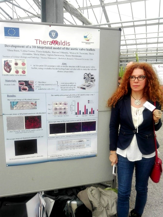
1Elena Butoi, 1Letitia Ciortan, 1Florin Iordache, 1Razvan D Macarie, 1Monica M. Tucureanu, 1Dorin Alexandru, 1Horia Maniu, 2Agneta Simionescu, 1Ileana Manduteanu
1Institute of Cellular Biology and Pathology “Nicolae Simionescu”, Bucharest, Romania
2Clemson University, USA
Introduction. Calcific aortic valve disease is a common heart valve disease in the modern world being a progressive process, with initial valvular endothelium inflammation, followed by fibrotic thickening and extensive calcification of the valve leaflets. Physiologically suitable in vitro models are required to study the disease progression and to develop potential therapeutic interventions.
Aim: to bio-print a 3D construct with a similar structure with the human aortic valve leaflet, using ECM-BioInkTM, human valvular cells and the platform 3DDiscovery (RegenHU, Switzerland)
Materials/Methods. Human fibroblasts, valvular interstitial cells (VICs) and endothelial cells (EC) were cultivated using standard cell culture methods. Fibroblasts or VICs were encapsulated in a bioink and bioprinted in 3 to 5 layers and EC were cultivated on the construct surface. The constructs were maintained in culture for 7-21 days. The stability and cell viability of printed 3D structures were followed in time, by MTT assay. The morphology of constructs was realized by hematoxylin-eozin and phalloidin staining. Cell phenotype was evaluated by Western Blot and immuno-histochemistry using specific anti-bodies.
Results/Discussions. In the current study, we implement 3D bioprinting to produce e structures similar to aortic human valves, with fibroblast/VIC encapsulated in a hydrogel and a EC layer on the top. Encapsulated fibroblasts or VICs developed a well-defined cell network within hydrogel and were viable over 21 days in culture. Encapsulated VICs expressed low levels of alpha-smooth muscle actin (α-SMA). Moreover, preliminary data obtained by Western blot analysis, indicate that bioprinted VICs have a less activated phenotype than those grown in bi-dimensional culture, and their maintaining for longer time in 3D culture favors the non-activated phenotype of VICs. Ongoing experiments to seed EC on the VIC construct are in progress.
Conclusions. Our results demonstrate the ability of the system to bio-print well 3D cell constructs with structure similar with the aortic valve leaflet with viable cells and phenotype retention over 21 days.
Acknowledgements. “This work is supported by the Competitiveness Operational Programme 2014-2020, Priority Axis1/Action 1.1.4/, Financing Contract no.115/13.09.2016/ MySMIS:104362“