2019
11TH NATIONAL CONGRESS WITH INTERNATIONAL PARTICIPATION AND 37TH ANNUAL SCIENTIFIC SESSION OF ROMANIAN SOCIETY FOR CELL BIOLOGY, June 20th-23rd, Constanta
Daniela Rebleanu1, Geanina Voicu1, Cristina Ana Constantinescu1, Letitia Ciortan1, Agneta Simionescu2, Ileana Manduteanu1, Manuela Calin1
1Institute of Cellular Biology and Pathology “Nicolae Simionescu” of Romanian Academy, Bucharest, Romania; 2Department of Bioengineering, Clemson University, United States of America
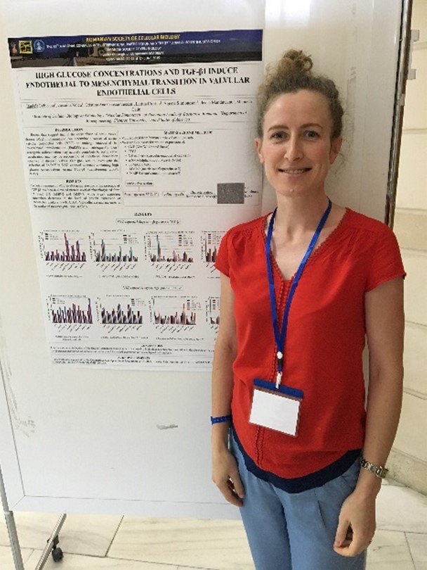
Introduction. Recent data suggest that in the early phase of aortic valve disease (AVD), inflammation can determine a subset of aortic valvular endothelial cells (VEC) to undergo endothelial to mesenchymal transformation (EndMT) and, subsequently their osteogenic differentiation may actively contribute to aortic valve calcification and may be responsible of endothelial denudation observed in diseased valves.
Purpose. To investigate the induction of EndMT in VEC exposed to media containing high glucose concentrations or/and TGF-β1 growth factor.
Methods. VEC were isolated from human aortic valves of patients who have undergone valve replacement and separated from VIC (valvular interstitial cells) using anti-CD31 magnetic beads. VEC were exposed to media containing normal (NG, 5.5 mM) or high glucose concentrations (HG, 25 mM or 30 mM) in the absence or the presence of TGF-β1 (10, 50, 100 ng/ml) for different periods of time (5 or 8 days). The medium was refreshed every two days. Then, VEC were lysed and analyzed by Western blot assay for endothelial cells markers (von Willebrand factor (vWF), CD 31, VE-cadherin), mesenchymal cell specific protein α- smooth muscle actin (SMA), and proteins that positively regulate the EndMT process (Smad2/3, MMP-2 and MMP-9).
Results. Five-day exposure of VEC to HG in the absence or the presence of TGF-β1 resulted in elevated protein level of phosphorylated form of Smad 2/3, MMP-2 and MMP-9, while 8-day exposure, determines decreases in the level of protein expression of endothelial markers (vWF, CD31, VE-cadherin) and increases in the marker of mesenchymal cells, α-SMA.
Conclusions. Our data show an induction of the EndMT transition process in VEC exposed to high glucose concentrations either in the absence or the presence of TGF-β1.
Acknowledgements. This work was supported by the Competitiveness Operational Programme 2014-2020, Priority Axis1/Action 1.1.4/, THERAVALDIS Project, contract no.115/13.09.2016/ MySMIS:104362.
Keywords: endothelial to mesenchymal transformation (EndMT), valvular endothelial cells, high glucose.

