2018
10TH NATIONAL CONGRESS WITH INTERNATIONAL PARTICIPATION AND 36TH ANNUAL SCIENTIFIC SESSION OF ROMANIAN SOCIETY FOR CELL BIOLOGY, June 6-9, Craiova
Monica Tucureanu1, Letitia Ciortan1, Razvan Macarie1, Mihaela Vadana1, Sergiu Cecoltan1, Elena Butoi1, Agneta Simionescu2, Ileana Manduteanu1
1Institute of Cellular Biology and Pathology “Nicolae Simionescu”, Bucharest, Romania
2Clemson University, USA
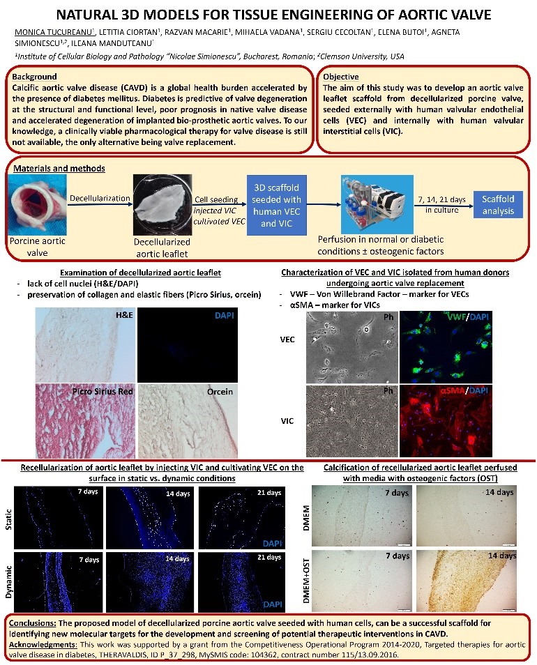
Calcific aortic valve disease (CAVD) is a global health burden accelerated by the presence of diabetes mellitus. Diabetes is predictive of valve degeneration at the structural and functional level, poor prognosis in native valve disease and accelerated degeneration of implanted bio-prosthetic aortic valves. To our knowledge, a clinically viable pharmacological therapy for valve disease is still not available, the only alternative being valve replacement. This address the need for the development of physiologically suitable in vitro models to study the valvular disease progression. The aim of this study was to develop an aortic valve leaflet scaffold from decellularized porcine valve, seeded externally with human valvular endothelial cells (VEC) and internally with human valvular interstitial cells (VIC). Fresh porcine aortic valves were collected and decellularized by immersion in different solutions (hypotonic solutions, detergents, deoxyribonuclease and ribonuclease). Decellularization efficiency and extracellular matrix integrity were evaluated by histological staining. Decellularized leaflets were seeded by injecting VICs in the scaffold matrix and cultivating VECs on the surface. The scaffolds were maintained in static or dynamic conditions for 7, 14 and 21 days in normal or diabetic conditions in the presence or absence of osteogenic factors. Immunohistochemistry analysis revealed the presence of VICs within the leaflet matrix, in a larger number in dynamic conditions compared with static conditions. VECs were organized as a well-defined monolayer at the scaffold surface in all experimental conditions. Moreover, we observed the calcification of valvular leaflets after 14 days of perfusion with media containing osteogenic factors, as determined by IHC. These results suggest that this scaffold of decellularized porcine aortic valve seeded with human cells, can be a successful model for identifying new molecular targets for the development and screening of potential therapeutic interventions in CAVD.
Keywords: aortic valve leaflet scaffold, recellularization, calcification.
Acknowledgments: This work was supported by a grant from the Competitiveness Operational Program 2014-2020, Targeted therapies for aortic valve disease in diabetes, THERAVALDIS, ID P_37_298, MySMIS code: 104362, contract number 115/13.09.2016.