2019
11TH NATIONAL CONGRESS WITH INTERNATIONAL PARTICIPATION AND 37TH ANNUAL SCIENTIFIC SESSION OF ROMANIAN SOCIETY FOR CELL BIOLOGY, June 20th-23rd, Constanta
Cristina Ana Constantinescu1, Daniela Rebleanu1, Geanina Voicu1, Letitia Ciortan, Sergiu Cecoltan, Dan Simionescu1,2, Agneta Simionescu1,2, Ileana Manduteanu1,
Manuela Calin1
1Institute of Cellular Biology and Pathology “Nicolae Simionescu”, Bucharest, Romania
2Department of Bioengineering, Clemson University, United States of America
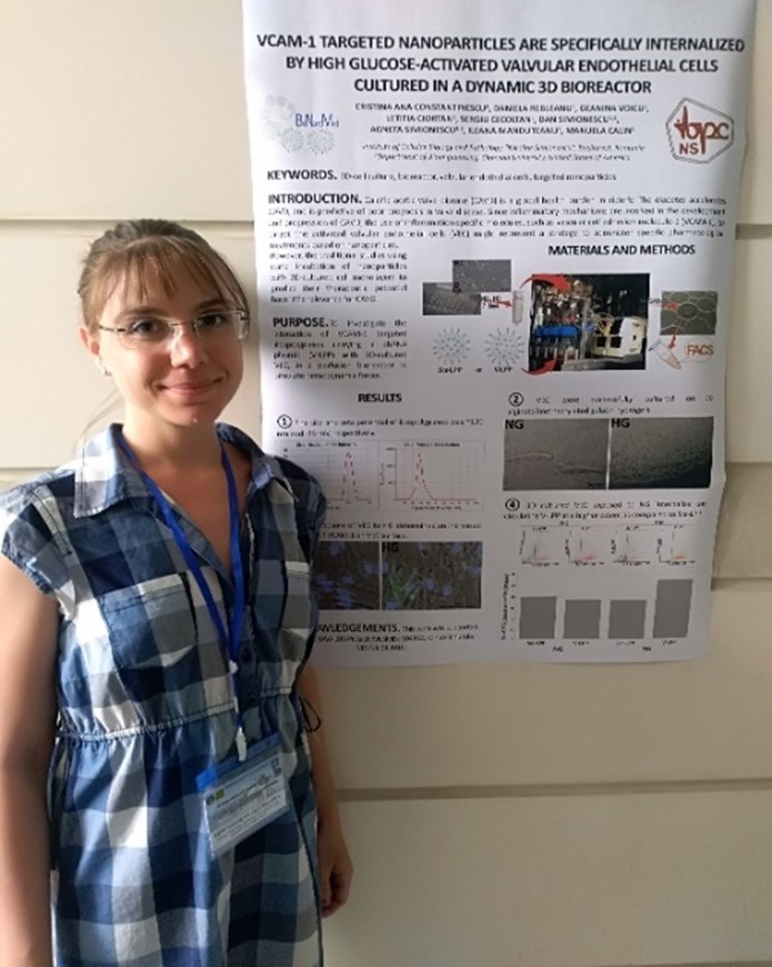
Introduction. Calcific aortic valve disease (CAVD) is a global health burden in elderly. The diabetes accelerates CAVD, and is predictive of poor prognosis in valve disease. Since inflammatory mechanisms are involved in the development and progression of CAVD, the use of inflammation-specific molecules, such as vascular cell adhesion molecule-1 (VCAM-1), to target the activated valvular endothelial cells (VEC) might represent a strategy to administer specific pharmacological treatments based on nanoparticles. However, the traditional studies using static incubation of nanoparticles with 2D-cultured cell monolayers to predict their therapeutic potential have little relevance for CAVD.
Purpose. To investigate the interaction of VCAM-1 targeted lipopolyplexes carrying a shRNA plasmid (V-LPP) with 3D-cultured VEC, in a perfusion bioreactor to simulate hemodynamic forces.
Materials and Methods. V-LPP were obtained by coupling a peptide recognizing VCAM-1 to anionic liposomes encapsulating polyplexes made of fullerene-polyethyleneimine (C60-PEI) and short hairpin (sh) RNA. VEC were cultured on 3D constructs consisting of alginate/methacrylated gelatin hydrogels. The 3D cultured VEC were exposed for five days to normal medium or to medium containing high-glucose concentrations (HG). Then, the 3D constructs were mounted in special designed supports in a 3D Perfusion Bioreactor™ (Biotek) and incubated for 24 hours with fluorescently-labeled lipopolyplexes non-targeted (Scr-LPP) or targeted to VCAM-1 (V-LPP). At the end of the incubation period, the VEC were detached and investigated by flow cytometry to follow the lipopolyplexes uptake.
Results and Discussions.
1) the size and zeta potential of lipopolyplexes was ~120 nm and -39 mV, respectively; 2) VEC were successfully cultured on 3D alginate/methacrylated gelatin hydrogels; 3) the exposure of VEC to HG determines an increased expression of VCAM-1 on VEC’ surface; 4) 3D cultured VEC exposed to HG internalize the circulating V-LPP at a higher extent as compared to Scr-LPP.
Acknowledgements. This work was supported by the THERAVALDIS Project: MySMIS: 104362, contract number 115/13.09.2016.
Keywords. 3D-cell culture, bioreactor, valvular endothelial cells, targeted nanoparticles.